 |
|||||||||||||||
go to
ARCHIVE
for topics on
all the days
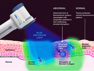
Here is the description by the company VELscope® Technology on the basic premise of tissue fluorescence visualization.
This tool “allows clinicians to see cellular, structural, and/or metabolic activity changes in oral mucosal tissues by observing the fluorescence response of oral tissues in response to light excitation.
Natural tissue fluorescence is caused by "fluorophores". When fluorophores are excited by light of an appropriate wavelength (for example, blue), they emit their own light at a longer wavelength (for example, green).
Abnormal fluorescence patterns aid the clinician in visualizing unhealthy mucosal tissue that sometimes
cannot be seen with the naked eye, such as viral, fungal and bacterial infections, inflammation from a
variety of causes (including lichen planus and other lichenoid reactions), squamous papillomas and
salivary gland tumours.”
citation: http://www.velscope.com
blue fluorescence visualization
The scope on the right is small enough to hold in ones hand and the beam of light circumference small enough to project into the dental patient's mouth. That's what happened to me. I was give special protective glasses when this procedure was being applied.
Yes, good news - as my illuminated mucosal tissue did not show any abnormal cell patterns as would be revealed by this beam of blue fluorescent light
However, there is a recent propesective study conducted to evaluate the using of the Velscope device - see information on right below the Velscope description. It makes a color splash but is it really needed.
.
Challenge:
source: [link]
The role of direct visual fluorescent examination (VELscope) in
routine screening for potentially malignant oral mucosal lesions.
– McNamara KK1, Martin BD, Evans EW, Kalmar JR.
Screening for Oral Squamous Cell Carcinoma
Oral squamous cell carcinoma is a common malignant process with a multifactorial etiology. The median age at diagnosis is 62 years, and the most commonly affected sites are the tongue and floor of the mouth.[1,2] Oral healthcare providers are continuously seeking additional methods of evaluating patients for precancerous and cancerous oral lesions.
Abstract
OBJECTIVE:
Direct visual fluorescent examination (DVFE) is a proposed adjunct to conventional oral examination (COE). We evaluate the benefit of DVFE in screening for potentially malignant mucosal lesions in a general population of patients presenting for dental care.
STUDY DESIGN:
A total of 130 patients were evaluated by COE followed by DVFE. Areas clinically suspicious by COE or with positive DVFE (visual fluorescence loss [VFL]) underwent surgical biopsy. Association between COE and DVFE was assessed and compared with histopathology.
RESULTS:
A total of 42 subjects had one or more areas of VFL, yet histologic evidence of premalignancy/malignancy was only identified in a single individual. Further, one lesion negative by DVFE exhibited epithelial dysplasia. DVFE was statistically different from scalpel biopsy (P = .0001). No difference was found between COE and scalpel biopsy (P = 1.0).
CONCLUSIONS:
Results suggest that COE is more valid than DVFE at discriminating benign mucosal alterations from premalignancy and do not support use of DVFE as an oral cancer screening adjunct.
For a copy of the Abstract Send to:
Oral Surg Oral Med Oral Pathol Oral Radiol. 2012 Nov;114(5):636-43. doi: 10.1016/j.oooo.2012.07.484.
The role of direct visual fluorescent examination (VELscope) in routine screening for potentially malignant oral mucosal lesions.
McNamara KK1, Martin BD, Evans EW, Kalmar JR.
Author information
Copyright © 2012 Elsevier Inc. All rights reserved.